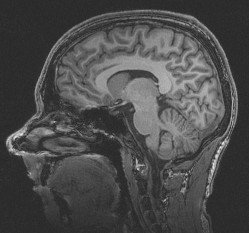
Magnetic resonance imaging (MRI) makes use of the magnetic properties of certain atomic nuclei. An example is the hydrogen nucleus (a single proton) present in water molecules, and therefore in all body tissues. The hydrogen nuclei behave like compass needles that are partially aligned by a strong magnetic field in the scanner.The nuclei can be rotated using radio waves, and they subsequently oscillate in the magnetic field while their magnetization returns to equilibrium. Simultaneously they emit a radio signal. This is detected using antennas (coils) and can be used for making detailed images of body tissues.
Unlike some other medical imaging techniques, MRI does not involve radioactivity or ionising radiation. The frequencies used (typically 40-130 MHz) are in the normal radiofrequency range, and there are no adverse health effects. Very detailed images can be made of soft tissues such as muscle and brain.The MR signal is sensitive to a broad range of influences, such as nuclear mobility, molecular structure, flow and diffusion. MRI is consequently a very flexible technique that provides measures of both structure and function.
Further information on MRI techniques
- YouTube video demonstrating the Magnetic Resonance phenomenon
- A more detailed explanation of MR including animations and software demonstrations.
- A widely used introduction to magnetic resonance techniques and other material (including simulators and animations) made in connection with open DRCMR courses.
- An introduction to the use of MR-Scanning (Danish)


