Brain structure determines function. Disentangling regional microstructural properties and understanding how these properties constitute brain function is a central goal of neuroimaging of the human brain and a key prerequisite for a mechanistic understanding of brain diseases and their treatment. Previous studies using magnetic resonance (MR) imaging have established links between regional brain microstructure and inter- individual variation in brain function, but this line of research has been limited by the non-specificity of MR-derived markers. This hampers the application of MR imaging as a tool to identify specific fingerprints of the underlying disease process.
In this project, we advance methodology on state-of-the-art MR hardware and harvest the synergy of these methods to realize Cell-specific in vivo MORPHometry (C-MORPH) of the intact human brain and spinal cord. In focus is the hybrid of advanced diffusion encoding strategies, spectroscopy and novel imaging approaches.
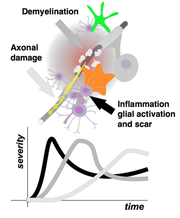
Our aim is to establish methods and analyses to derive cell-type specific tissue properties in the healthy and diseased brain. In particular, we aim for isolating the severity of specific pathological processes and their development over time. To date, such processes are more or less superimposed in conventional clinical imaging data. Once validated, the experimental methods and analyses will be simplified and adapted to provide clinically applicable tools. This will push the frontiers of MR-based personalized medicine, guiding therapeutic decisions by providing sensitive probes of cell-specific microstructural changes caused by inflammation, neurodegeneration or treatment response.
The C-MORPH project was initiated in the end of 2018 with the ERC Staring Grant awarded to the P.I. Henrik Lundell.
