Headed by Professor Hartwig Siebner, the aim of the Neuroimaging in Multiple Sclerosis (NiMS) group is to push the frontiers of MRI to capture MS-related tissue damage and uncover the underlying pathophysiological mechanisms. The group uses a wide range of MR-based techniques such as functional MRI, magnetic resonance spectroscopy (MRS), and diffusion weighted imaging (DWI), but also electrophysiological methods such as electroencephalography (EEG) and transcranial magnetic stimulation (TMS). The current focus is on exploiting the potential of MRI at ultra-high field strength (7 tesla) to detect neocortical involvement and characterize microstructural changes in cerebral white matter.

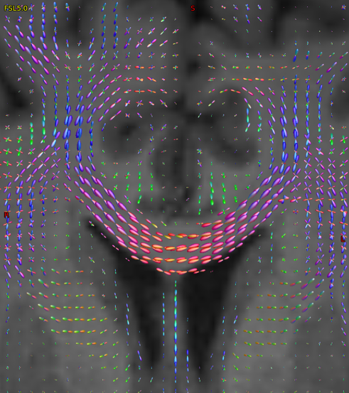
An image based on diffusion weighted imaging demonstrating fibre directionality in the coronal representation of corpus callosum. Estimating voxelwise fibre directions is the first stage in the process of mapping structural brain connectivity in patients with multiple sclerosis.
Neuroimaging research in the field of MS has a long tradition at DRCMR and greatly benefits from a long-standing and inspiring collaboration with the Danish Multiple Sclerosis Centre, Department of Neurology, Copenhagen University Hospital Rigshospitalet, Denmark. In recent years, the NiMS group pursued several lines of neuroimaging research, including studies of the blood-brain barrier dysfunction in normal appearing white matter (PhD project by Henrik Lund) and the atrophy pattern of the upper cervical spinal cord (Ellen Grade and Henrik Lundell). Together with the DRCMR Reader Centre, the NIMS group participates in the analysis of the MRI data that are acquired at DRCMR as part of investigator-driven as well as company initiated clinical trials.
An overarching theme of our research is to examine how MS alters functional and structural brain connectivity and how such MS-related connectivity changes contribute to clinical disability (Kasper W. Andersen, Kristoffer H. Madsen). Using rs-fMRI, the NiMS group was able to identify distinct changes in functional connectivity in the motor resting-state network in a group of patients with relapsing-remitting or secondary progressive MS (PhD project by Anne-Marie Dogonowski). We found that MS impairs regional functional connectivity in the cerebellum. At the brain network level, patients with MS showed a more widespread coupling of the basal ganglia with the motor resting-state network, indicating an impaired “funneling” function of the basal ganglia in MS. Moreover, resting-state connectivity of pre-motor cortex reflected the degree of disability on the Expanded Disability Status Scale (EDSS).
Key projects
What causes fatigue in patients with multiple sclerosis? – A network perspective
We are currently conducting a comprehensive multi-modal brain mapping study, in which we aim at delineating abnormalities in brain function and structure that lead to fatigue in patients with MS. Fatigue is one of the most common symptoms in Multiple Sclerosis (MS). It has been suggested that fatigue is a consequence of disease-related microstructural alterations in specific white matter (WM) tracts. By applying anatomical connectivity mapping (ACM), we wish to probe whether fatigued MS patients have different levels of anatomical connectedness compared to non-fatigued MS patients. (PhD projects by Olivia Svolgaard and Christian Bauer).
Cortical lesion in primary sensorymotor hand area and their impact on dexterity in multiple sclerosis: a 7T MRI study
In this project, we wish to clarify the impact of regional cortical lesions within the sensorymotor hand area (SM1-HAND) on cortical function and manual dexterity. Exploiting the increased sensitivity of ultra-high field 7 Tesla MRI to detect cortical lesions, we will assess the number, size and regional distribution of cortical lesions in SM1-HAND, and relate regional lesion load in SM1-HAND to MRI-based, electrophysiological, and behavioural correlates of hand function (PhD project by Mads Alexander Just Madsen).
Diffusion weighted magnetic resonance spectroscopy at ultra-high field:
Unravelling microstructural changes in cerebral white matter in patients with multiple sclerosis
We are currently pursuing the first clinical ultra-high field (7T) MR study in Denmark. In this project, we will combine magnetic resonance spectroscopy with diffusion MRI to shed new light into the microstructural alterations in major motor white-matter tract caused by MS. The project is conducted by Senior Researcher Henrik Lundell who received a “Sapere Aude” award by the Danish Council for Independent Research.
Research funding:
We wish to thank the Danish Multiple Sclerosis Society, the Danish Council for Independent Research, Jascha fonden, Torben Fogs og Erik Triers Fond, and Biogen-Idec, Denmark, for their generous support. Christian Bauer´s PhD project was co-financed by Metropolitan University College, Copenhagen, Denmark.


