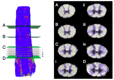
 This high-resolution study was performed to understand the underlying anisotropy observed in in vivo diffusion-weighted datasets of the human spinal cord. Fine structures in both the white-matter and grey-matter are visible. One important feature is the collateral fibers forming fiber crossings in plane, perpendicular to the main descending/ascending fibers. Those are shown in blue in the figure below to the right. White matter in the spinal cord is indeed not axons in only one direction!
Facts about the dataset:
Sequence: diffusion weighted spin echo with single line readout. Image matrix: 64x64, Voxel size: axial resolution 188x188 µm^2, slice thickness 375 µm, slices: 70, TR= 4500 ms, TE= 50 ms. G = 180 mT/m, small delta: 8 ms, big delta: 20 ms, maximum b-value 4525 s/mm^2, 105 directions and 25xb0. 4 repetitions. The sample covers the cervical spinal cord of a vervet monkey. Each repeated scan contain a 4D dataset in NIFTI format where 3D image volumes are ordred as: First all b0 image volumes followed by all the diffusion weighted image volumes.
To cite:
Lundell H, Nilsen JB, Ptito M, Dyrby TB, Distribution of collateral fibers in the monkey cervical spinal cord detected with diffusion weighted magnetic resonance imaging, Neuroimage, 2011, 56(3):923-9
Download the ex vivo spinal cord dataset and Camino gradient directions here
Observed problems or artifacts: none
Funding:
Elsass Foundation