Cortical lessions and grey matter dysfunction in Multiple Sclerosis
Imaging plays a pivotal role in multiple sclerosis (MS) diagnosis and monitoring. It can help us uncover and understand the yet unknown mechanisms that contribute to the large variety of symptoms in MS.
For example, cortical pathology is now known to significantly contribute to disability in multiple sclerosis (MS) and has therefore attracted considerable interest in the last decade. However, we are yet to understand the impact of cortical damage on both the connectivity and functional integrity of the affected area and other parts of the central nervous system.
In this project, we exploit the greater sensitivity of 7 Tesla magnetic resonance imaging (MRI) to cortical lesions, together with quantitative imaging sequences and transcranial magnetic stimulation (TMS) to comprehensively map the role of cortical damage in different subtypes of MS.
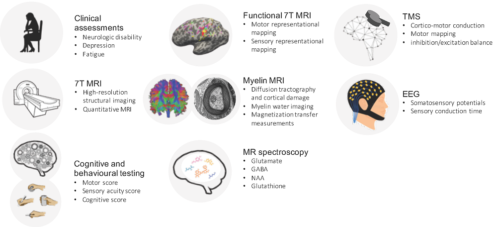
Overview of the experimental protocol and methodology used in the project.
Using both MRI and TMS, this project addresses two main research questions:
- 1)What is the impact of individual cortical lesions, specifically in the primary sensorimotor hand area, on hand dexterity in different subtypes of MS
- 2) What role do focal and diffuse cortical myelin changes play in relation to physical and cognitive function in primary progressive MS
In close collaboration with the Danish Multiple Sclerosis Center at Rigshospitalet Glostrup, we are in the unique position to investigate these questions in MS patients at all stages of the disease.
Status of the project
Our preliminary results show that a cortical lesion in the primary sensorimotor hand area significantly reduces both manual dexterity and sensory acuity of the fingers. Additionally, our results demonstrate that TMS is sensitive to the disruption of cortical function due to cortical lesions, and that this disruption might be related to increased disability.
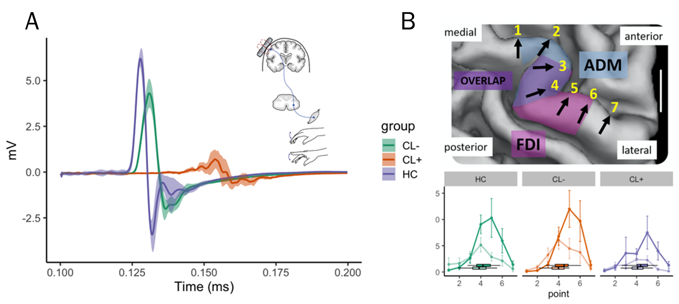
(A) The three motor evoked potentials (MEP) traces show that the onset latency was longer and lower in amplitude for both relapsing remitting MS patients compared to a healthy control (HC), but much more so in the patient with cortical damage (CL+). (B) TMS motor mapping successfully separates the representation of the FDI (bold) and ADM (faded color) muscle in all three groups.
Impact
This project will reveal key insights into how cortical demyelination and damage, both regionally and globally, contribute to cognition and motor impairment in MS, two major disabling problems for patients. We will advance the possibilities of MRI to capture cortical involvement in Danish MS patients with the goal to improve individual stratification, monitoring of disease progression and capturing of the individual response to therapy.
Funding Sources
Scleroseforeningen, Danmarks Frie Forskningsfond, Hvidovre Hospital, Gangsted fonden og Lundbeckfonden.
![]()
![]()
![]()