DRCMR Logo 300px Color
 

Neurophysics


The Neurophysics group is headed by Professor Axel Thielscher and situated both at DRCMR and DTU Health Tech. Our main focus is on advancing non-invasive transcranial brain stimulation (TBS) methods as a means to modulate and shape brain activity. Most TBS approaches use electric currents that are focally induced in superficial brain areas. We develop and apply biophysical models to reveal and optimize the current flow patterns in the brain and to estimate their impact on neural activity. The computational modeling work is complemented by applying neuroimaging approaches such as functional MRI (fMRI) and electroencephalography (EEG) to better characterize the impact of neurostimulation on brain activity. We are interested in human sensorimotor integration and motor control, which sets the neuroscientific scene in which we employ and test the NTBS methods.

The Neurophysics group contributes to a range of interdisciplinary project at DRCMR, such as the former BASICS project. We have tight interactions with the Control of Movement and the Brain Network Modulation groups (headed by Hartwig Siebner), the Computational Neuroimaging group (headed by Kristoffer H. Madsen) and the MR Acquisition Technology group (headed by Lars G. Hanson).

Funding sources

We would like to thank our funding sources.

Lundbeck fondenNovo Nordisk fondenSDC  STIPED Innovation Fund Danmark


Key projects


Simulation of Non-invasive Brain Stimulation (SimNIBS)

 Neurophysics Figure 1


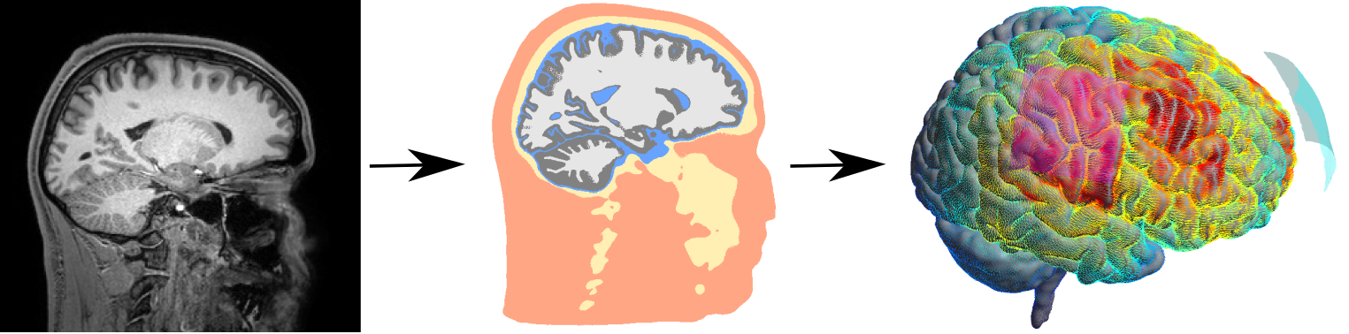
SimNIBS is both a scientific and an open-source software development project (Thielscher et al. 2011). We use field calculations to estimate the current flow generated by TBS methods in realistic head models, which are reconstructed from structural MR images. This allows us to determine the likely stimulated brain areas, and in turn to optimize the stimulation patterns. After developing the methodology, we are currently focusing on testing how well the estimated fields correlate with the experimentally observed stimulation effects. Simultaneously, we are strongly extending SimNIBS to include optimization methods for multi-electrode current steering for transcranial electric stimulation (TES) and to enable forward calculations for EEG in highly realistic head models. Oula Puonti is the main contributor to this project.

Automatic Whole-Head Segmentation
Neurophysics Figure 2

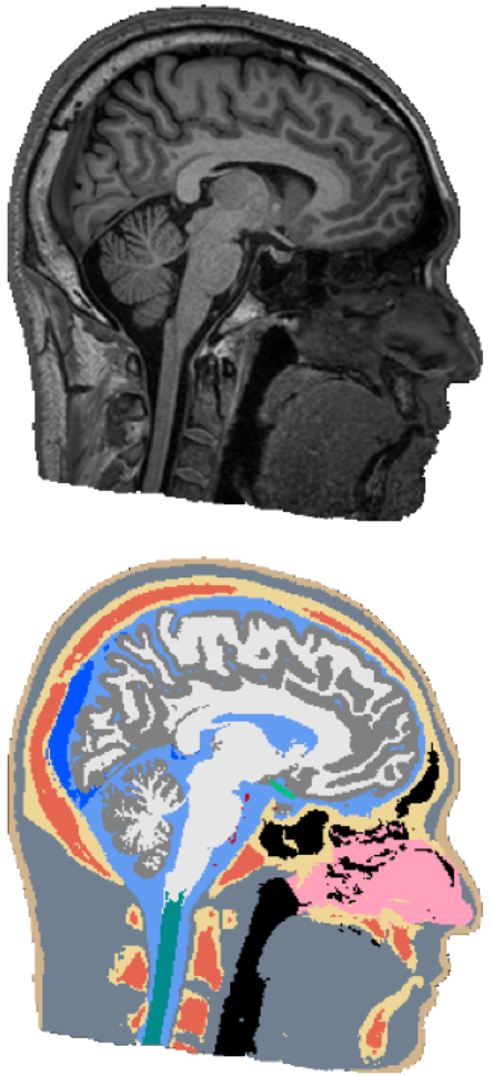
The electric fields induced by TBS are shaped largely by the individual anatomy of a target subject, and have complex and often counter-intuitive spatial distribution. In order to do targeted stimulation, or to locate sources in EEG or MEG, access to accurate models of the head anatomy is crucial. We are currently working on an automated segmentation approach for robust and accurate segmentation of 14 different head tissues from a subject’s MRI scan. The approach should generalize across different MRI scanners and scan-sequences, as well as work robustly on clinical MRI data, which often has a low resolution and movement-related artifacts. In the future, we are also planning to include automatic segmentation of pathologies, such as lesions or tumors, to facilitate the use of NIBS in clinical applications.Oula Puonti is the main contributor to this project.

Advanced Methods for MREIT and MRCDI

Neurophysics Figure 3
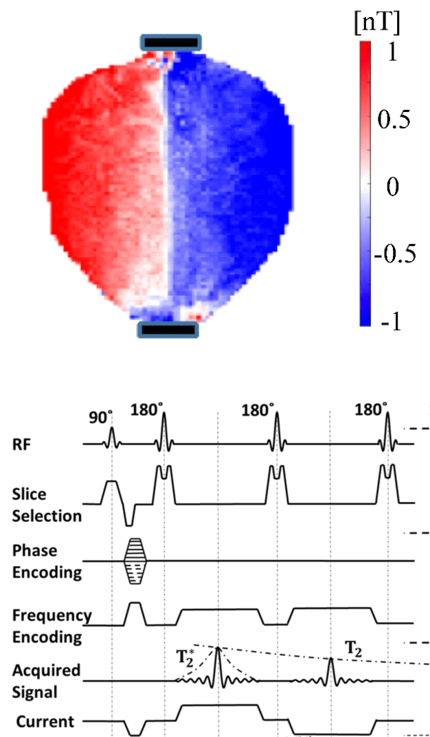
Magnetic resonance electrical impedance tomography (MREIT) and magnetic resonance current density imaging (MRCDI) are evolvling as methods to directly measure the ohmic conductivity distribution in the head and the current flow pattern generated by transcranial electric stimulation (TES). They would be tremendously useful to validate the field estimates from computational models, or might be used as alternative to them giving similar information. A main challenge for establishing MRCDI and MREIT in humans is the limited sensitivity of the MR measurements. We have demonstrated the so far highest sensitivity to injected currents worldwide (Göksu et al. 2018), and have shown reliable mappings of magnetic fields generated by injected currents as low as 1 milliampere. Currently, we are focusing on using the measurements to improve the SimNIBS forward simulations, and to mature the MR methods for use in larger studies. Cihan Göksu and Frodi Gregersen are main contributors to this project.

Weak Transcranial Focused Ultrasound Stimulation (TFUS)  Neurophysics Figure 4

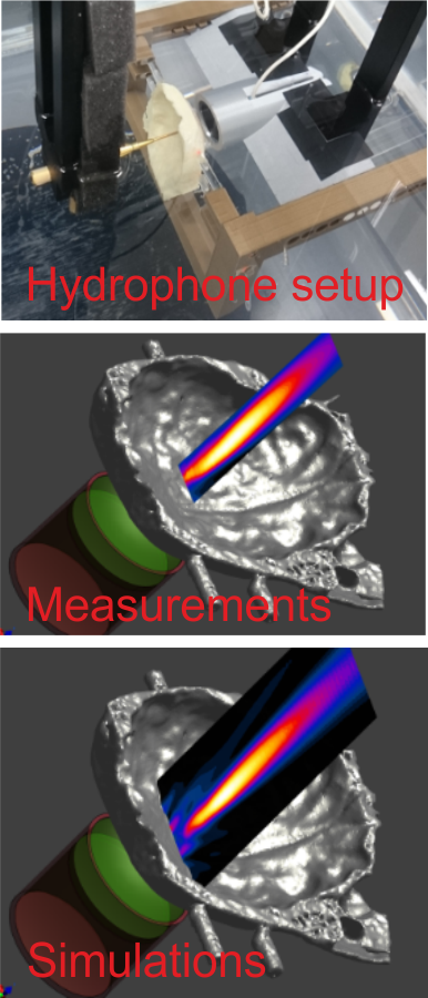
TFUS is emerging as new stimulation method with superior spatial resolution and the possibility to reach deeper areas of the brain. The existing data points towards a favorable safety profile of TFUS and suggests that it might be well usable in human applications (Pasquinelli et al. 2019). Computational dosimetry approaches will have a key role in future TFUS applications in order to ensure safe and neurally effective ultrasound intensities in the brain. However, dose control is made challenging by the strong impact of the heterogeneous skull on the TFUS beam, which can be difficult to simulate accurately. For this reason, we are working on the validation of simulations that are based on individual CT data of the skull. The simulations are based on the Sim4Life platform in a collaboration with the IT’IS foundation. 

Characterizing the Effects and Side-effects of Transcranial Magnetic StimulationNeurophysics Figure 5

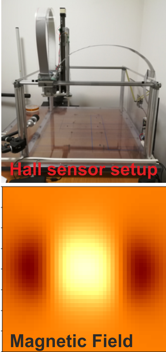
Transcranial magnetic stimulation (TMS) is a non-invasive method to stimulate cortical regions of the brain. It has established clinical and research applications, such as the treatment of depression. While TMS has been demonstrated to be effective, it causes also unwanted side-effects like pain and muscle twitches. We will develop models of existing commercial coils that will serve as an input to SimNIBS and extend our volume conductor models of the head in order estimate the stimulation of peripheral nerves in the skin. Our aim is to develop a quantitative computational model to estimate both the desired stimulation effect in the brain and the side-effects of TMS. We envision that this will open up new ways to develop more comfortable and accurate TMS coils. Maria Drakaki, Industrial PhD student at DTU and MagVenture, is the main contributor to this project. 

Integrating TBS with Neuroimaging Neurophysics Figure 6

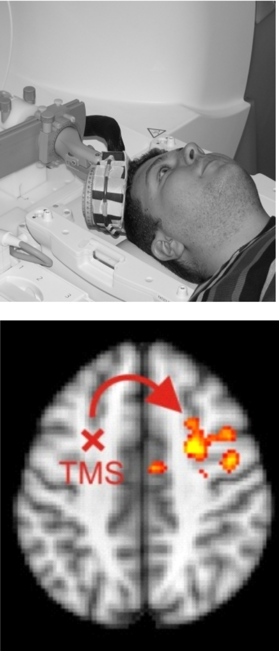
We are combining TBS with functional MRI (fMRI) to shed light on the stimulation effects at the brain network level and to characterize how the neural TBS effects depend on the physical TBS fields and the brain state. For example, we have shown that controlling the brain state by a behavioral task modulates the TBS effects, which could be used for optimizing TBS interventions (Moisa et al. 2012). Currently, in collaborations with the Brain Network Modulation and Computational Neuroimaging groups, we are integrating MR-based imaging of the neural TBS effects with SimNIBS calculations and MRCDI measurements of the induced fields. Our aim is to assess the usefulness of computational dosimetry approaches for predicting and optimizing physiological stimulation effects.

Novel methods for measuring neural magnetic fields Neurophysics Figure 7

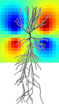
Nitrogen-vacancy centers in diamond are emerging as new sensors for highly sensitive magnetometry with superior spatial resolution and the ability to work at ambient temperatures. We are contributing to the Interdisciplinary Synergy Project “Interfacing emerging quantum technology with biology and neurophysiology” (BioQ) that is funded by Novo Nordisk Fonden and driven by our collaborators Ulrik Lund Andersen and Alexander Huck (DTU Physics). Extending our work in the EXMAD project, we aim to enable highly sensitive measurements of neural magnetic fields of neurons both in vitro and in humans.


Selected Publications

Webb JL, Troise L, Hansen NW, Olsson C, Wojciechowski AM, Achard J, Brinza O, Staacke R, Kieschnick M, Meijer J, Thielscher A, Perrier J-F, Berg-Sørensen K, Huck A, Andersen UL. 2021. Detection of biological signals from a live mammalian muscle using an early stage diamond quantum sensor. Scientific Reports. 11(1):1-11. https://doi.org/10.1038/s41598-021-81828-x

von Conta J, Kasten FH, Ćurčić-Blake B, Aleman A, Thielscher A, Herrmann CS. 2021. Interindividual variability of electric fields during transcranial temporal interference stimulation (tTIS). Scientific Reports. 11(1):1-12. https://doi.org/10.1038/s41598-021-99749-0

Splittgerber M, Borzikowsky C, Salvador R, Puonti O, Papadimitriou K, Merschformann C, Biagi MC, Stenner T, Brauer H, Breitling-Ziegler C, Prehn-Kristensen A, Krauel K, Ruffini G, Pedersen A, Nees F, Thielscher A, Dempfle A, Siniatchkin M, Moliadze V. 2021. Multichannel anodal tDCS over the left dorsolateral prefrontal cortex in a paediatric population. Scientific Reports. 11(1):1-15. https://doi.org/10.1038/s41598-021-00933-z

Shirinpour S, Mantell K, Li X, Puonti O, Madsen K, Haigh Z, Casillo EC, Alekseichuk I, Hendrickson T, Xu T. 2021. New tools for computational modeling of non-invasive brain stimulation in SimNIBS. Brain Stimulation: Basic, Translational, and Clinical Research in Neuromodulation. 14(6):1644. https://doi.org/10.1016/j.brs.2021.10.180

Saturnino GB, Madsen KH, Thielscher A. 2021. Optimizing the electric field strength in multiple targets for multichannel transcranial electric stimulation. Journal of Neural Engineering. 18(1): Article 014001. https://doi.org/10.1088/1741-2552/abca15

Numssen O, Zier A-L, Thielscher A, Hartwigsen G, Knösche TR, Weise K. 2021. Efficient high-resolution TMS mapping of the human motor cortex by nonlinear regression. NeuroImage. 245:1-11. https://doi.org/10.1016/j.neuroimage.2021.118654

Montanaro H, Pasquinelli C, Lee HJ, Kim H, Siebner HR, Kuster N, Thielscher A, Neufeld E. 2021. The impact of CT image parameters and skull heterogeneity modeling on the accuracy of transcranial focused ultrasound simulations. Journal of Neural Engineering. 18(4):1-28. https://doi.org/10.1088/1741-2552/abf68d

Mezger E, Rauchmann B-S, Brunoni AR, Bulubas L, Thielscher A, Werle J, Mortazavi M, Karali T, Stöcklein S, Ertl-Wagner B, Goerigk S, Padberg F, Keeser D. 2021. Effects of bifrontal transcranial direct current stimulation on brain glutamate levels and resting state connectivity: multimodal MRI data for the cathodal stimulation site. European Archives of Psychiatry and Clinical Neuroscience. 271(1):111-122. https://doi.org/10.1007/s00406-020-01177-0

Karadas M, Olsson C, Winther Hansen N, Perrier J-F, Webb JL, Huck A, Andersen UL, Thielscher A. 2021. In-vitro Recordings of Neural Magnetic Activity From the Auditory Brainstem Using Color Centers in Diamond: A Simulation Study. Frontiers in Neuroscience. 15:1-17. https://doi.org/10.3389/fnins.2021.643614

Gregersen F, Göksu C, Schaefers G, Xue R, Thielscher A, Hanson LG. 2021. Safety Evaluation of a New Setup for Transcranial Electric Stimulation during Magnetic Resonance Imaging. Brain Stimulation. 14(3):488-497. https://doi.org/10.1016/j.brs.2021.02.019

Göksu C, Scheffler K, Gregersen F, Eroğlu HH, Heule R, Siebner HR, Hanson LG, Thielscher A. 2021. Sensitivity and resolution improvement for in vivo magnetic resonance current-density imaging of the human brain. Magnetic Resonance in Medicine. 86(6):3131-3146. https://doi.org/10.1002/mrm.28944

Eroğlu HH, Puonti O, Göksu C, Gregersen F, Siebner HR, Hanson LG, Thielscher A. 2021. On the reconstruction of magnetic resonance current density images of the human brain: Pitfalls and perspectives. NeuroImage. 243:1-15. https://doi.org/10.1016/j.neuroimage.2021.118517

Dubbioso R, Madsen KH, Thielscher A, Siebner HR. 2021. The myelin content of the human precentral hand knob reflects interindividual differences in manual motor control at the physiological and behavioral level. The Journal of Neuroscience: the official journal of the Society for Neuroscience. 41(14):3163-3179. https://doi.org/10.1523/JNEUROSCI.0390-20.2021

Antonenko D, Grittner U, Puonti O, Flöel A, Thielscher A. 2021. Estimation of individually induced e-field strength during transcranial electric stimulation using the head circumference. Brain Stimulation. 14(5):1055-1058. https://doi.org/10.1016/j.brs.2021.07.001

Antonenko D, Grittner U, Saturnino G, Nierhaus T, Thielscher A, Flöel A. 2021. Inter-individual and age-dependent variability in simulated electric fields induced by conventional transcranial electrical stimulation. NeuroImage. 224:1-9. https://doi.org/10.1016/j.neuroimage.2020.117413

Weise K, Numssen O, Thielscher A, Hartwigsen G, Knösche TR. 2020. A novel approach to localize cortical TMS effects. NeuroImage. 209:1-17. Available from: 10.1016/j.neuroimage.2019.116486

Puonti O, Van Leemput K, Saturnino GB, Siebner HR, Madsen KH, Thielscher A. 2020. Accurate and robust whole-head segmentation from magnetic resonance images for individualized head modeling. NeuroImage. 219:1-17. Available from: 10.1016/j.neuroimage.2020.117044

Puonti O, Saturnino GB, Madsen KH, Thielscher A. 2020. Value and limitations of intracranial recordings for validating electric field modeling for transcranial brain stimulation. NeuroImage. 208:1-14. Available from: 10.1016/j.neuroimage.2019.116431

Pasquinelli C, Montanaro H, Lee HJ, Hanson LG, Kim H, Kuster N, Siebner HR, Neufeld E, Thielscher A. 2020. Transducer modeling for accurate acoustic simulations of transcranial focused ultrasound stimulation. Journal of Neural Engineering. 17(4):1-22. Available from: 10.1088/1741-2552/ab98dc

Habich A, Fehér KD, Antonenko D, Boraxbekk C-J, Flöel A, Nissen C, Siebner HR, Thielscher A, Klöppel S. 2020. Stimulating aged brains with transcranial direct current stimulation: Opportunities and challenges: Opportunities and challenges. Psychiatry Research - Neuroimaging. 306:1-9. Available from: 10.1016/j.pscychresns.2020.111179

Boayue NM, Csifcsák G, Aslaksen P, Turi Z, Antal A, Groot J, Hawkins GE, Forstmann B, Opitz A, Thielscher A, Mittner M. 2020. Increasing propensity to mind-wander by transcranial direct current stimulation? A registered report. European Journal of Neuroscience. 51(3):755-780. Available from: 10.1111/ejn.14347

Bikson M, Hanlon CA, Woods AJ, Gillick BT, Charvet L, Lamm C, Madeo G, Holczer A, Almeida J, Antal A, Ay MR, Baeken C, Blumberger DM, Campanella S, Camprodon J, Christiansen L, Colleen L, Crinion J, Fitzgerald P, Gallimberti L, Ghobadi-Azbari P, Ghodratitoostani I, Grabner R, Hartwigsen G, Hirata A, Kirton A, Knotkova H, Krupitsky E, Marangolo P, Nakamura-Palacios EM, Potok W, Praharaj SK, Ruff CC, Schlaug G, Siebner HR, Stagg CJ, Thielscher A, Wenderoth N, Yuan T-F, Zhang X, Ekhtiari H. 2020. Guidelines for TMS/tES Clinical Services and Research through the COVID-19 Pandemic. Brain Stimulation. 13(4):1124-1149. Available from: 10.1016/j.brs.2020.05.010

Saturnino GB, Siebner HR, Thielscher A, Madsen KH Accessibility of cortical regions to focal TES: Dependence on spatial position, safety, and practical constraints. Neuroimage. 2019 doi: 10.1016/j.neuroimage.2019.116183

Saturnino GB, Madsen KH, Thielscher A Electric field simulations for transcranial brain stimulation using FEM: an efficient implementation and error analysis. J Neural Eng. doi: 10.1088/1741-2552/ab41ba, 2019

Pasquinelli C, Hanson LG, Siebner HR, Lee HJ, Thielscher A Safety of transcranial focused ultrasound stimulation: A systematic review of the state of knowledge from both human and animal studies Brain Stimul. doi: 10.1016/j.brs.2019.07.024, 2019

Korshøj AR, Sørensen JCH, von Oettingen G, Poulsen FR, Thielscher A Optimization of tumor treating fields using singular value decomposition and minimization of field anisotropy. Phys Med Biol. 64(4):04NT03. 2019

Saturnino GB, Thielscher A, Madsen KH, Knösche TR, Weise K. A principled approach to conductivity uncertainty analysis in electric field calculations. Neuroimage. 188:821-834, 2019

Karadas M, Wojciechowski AM, Huck A, Dalby NO, Andersen UL, Thielscher A. Feasibility and resolution limits of opto-magnetic imaging of neural network activity in brain slices using color centers in diamond. Sci Rep. 8(1):4503, 2018.

Nielsen JD, Madsen KH, Puonti O, Siebner HR, Bauer C, Madsen CG, Saturnino GB, Thielscher A Automatic skull segmentation from MR images for realistic volume conductor models of the head: Assessment of the state-of-the-art. Neuroimage. 174:587-598, 2018.

Göksu, C., Hanson, L. G., Siebner, H. R., Ehses, P., Scheffler, K. & Thielscher, A. Human in-vivo brain magnetic resonance current density imaging (MRCDI). NeuroImage. 171, p. 26-39, 2018.

Göksu C, Scheffler K, Ehses P, Hanson L.G, Thielscher A. Sensitivity Analysis of Magnetic Field Measurements for Magnetic Resonance Electrical Impedance Tomography (MREIT), Magnetic Resonance in Medicine. 79, p. 748-760, 2018.

Bungert, A., Antunes, A., Espenhahn, S. & Thielscher, A. Where does TMS Stimulate the Motor Cortex? Combining Electrophysiological Measurements and Realistic Field Estimates to Reveal the Affected Cortex Position. Cerebral cortex, 27(11):5083-5094, 2017.

Minjoli, S., Saturnino, G. B., Blicher, J. U., Stagg, C. J., Siebner, H. R., Antunes, A. & Thielscher, A. The impact of large structural brain changes in chronic stroke patients on the electric field caused by transcranial brain stimulation. NeuroImage. Clinical. 15, p. 106-117, 2017.

Saturnino, G. B., Madsen, K. H., Siebner, H. R. & Thielscher, A. How to target inter-regional phase synchronization with dual-site Transcranial Alternating Current Stimulation.
NeuroImage. 163, p. 68-80, 2017.

Madsen, K.H., Ewald, L., Siebner, H.R., Thielscher, A. Transcranial Magnetic Stimulation: An Automated Procedure to Obtain Coil-specific Models for Field Calculations. Brain Stimulation 8, 1205-1208, 2015

Saturnino, G.B., Antunes, A., Thielscher, A. On the importance of electrode parameters for shaping electric field patterns generated by tDCS. Neuroimage 120, 25-35, 2015.

Moisa, M., Siebner, H.R., Pohmann, R., Thielscher, A. Uncovering a context-specific connectional fingerprint of human dorsal premotor cortex. J Neurosci 32, 7244-7252, 2012.

Thielscher, A., Opitz, A., Windhoff, M. Impact of the gyral geometry on the electric field induced by transcranial magnetic stimulation. Neuroimage 54, 234-243, 2011

Group Members

Axel Thielscher

Group Leader

Oula Puonti

Marie Louise Liu

Show all group members (10)

External Collaborators

Professor Ulrik Lund Andersen 

DTU Physics


Assoc. Professor Alexander Huck

DTU Physics


Assoc. Prof. Koen van Lempuut

Harvard University & DTU Compute


Assoc. Prof. Gottfried Schlaug

Beth Israel Deaconess Medical Center


Prof. Dr. Klaus Scheffler

Max Planck Institut for Biological Cybernetics


Assistant Prof. Hyunjoo Jenny Lee

KAIST


Professor Michael Siniatchkin

University of Bielefeld