By altering neuronal processing in the stimulated area NTBS can be used to characterize the causal and functional contribution of the stimulated cortex to a specific brain function and its functional interactions. The effects of NTBS can outlast the time of stimulation. Therefore, NTBS is widely used to induce plasticity in targeted brain circuits, bearing great scientific and therapeutic potential. Below are two examples of studies performed in the context of this workpackage.
Study 1
Raffin E, Pellegrino G, Di Lazzaro V, Thielscher A, Siebner HR (2015) Bringing transcranial mapping into shape: Sulcus-aligned mapping captures motor somatotopy in human primary motor hand area. NeuroImage 120:164–175.
In this work, we introduce and validate a novel linear TMS mapping approach of muscle-specific corticomotor representations. The approach can be used to obtain mediolateral spatial profiles of corticomotor excitability for individual intrinsic hand muscles and to trace state-dependant spatial shifts of these excitability profiles (e.g. tonic contraction versus rest) in the hand area of the human primary motor cortex M1HAND.
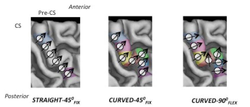
We obtained mediolateral corticomotor excitability profiles of the abductor digiti minimi (ADM) and first dorsal interosseus (FDI) muscles. We stimulated six targets located on a straight mediolateral line (STRAIGHT-450FIX) or seven targets in the posterior part of the crown of the central sulcus following the bending of the central sulcus (CURVED). CURVED mapping employed a fixed (CURVED-450FIX) or flexible coil orientation (CURVED-900FLEX). During relaxation, CURVED but not STRAIGHT mapping revealed distinct corticomotor excitability peaks in M1HAND. This mediolateral somatotopy was still present during tonic contraction of the ADM or FDI.
 |
 |
|
This figure illustrates the stimulation sites of STRAIGHT-450FIX (left panel), |
This figure illustrates the stimulation sites of STRAIGHT-450FIX (left panel), CURVED-450FIX and CURVED-900FLEX (left and middle panels). |
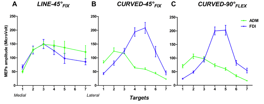
Linear medio-lateral profiles (i.e. distribution of the MEPs target by target) of the 2 muscles for the three mapping approaches. Only “CURVED-450FIX” and “CURVED-900FLEX” demonstrated a clear somatotopy of the FDI and ADM muscle representations. The MEPs amplitudes for ADM peaked in the medial portion of M1HAND whereas the MEPs amplitudes of the FDI muscle were maximal in the lateral part of M1HAND
Study 2
Delvendahl I, Lindemann H, Jung NH, Pechmann A, Siebner HR, Mall V (2014) Influence of Waveform and Current Direction on Short-Interval Intracortical Facilitation: A Paired-Pulse TMS Study. Brain Stimulat 7:49–58.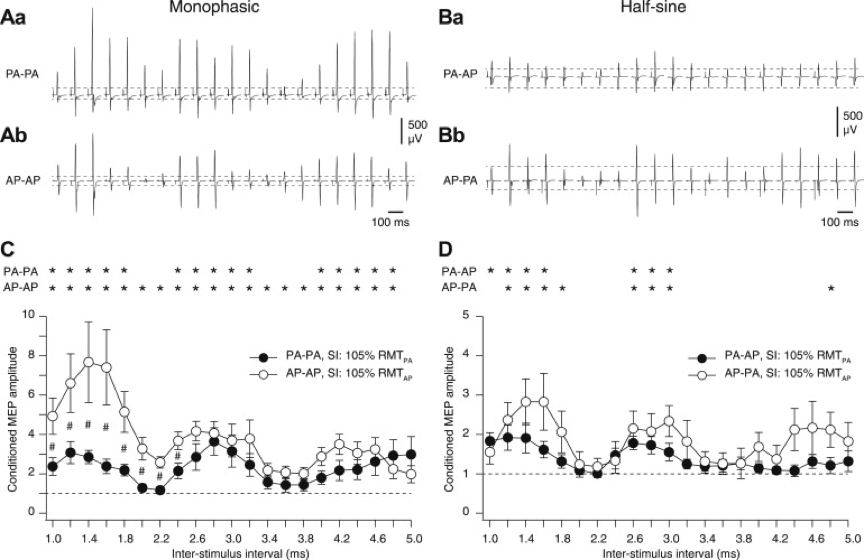
Transcranial magnetic stimulation of the human primary motor hand area can produce multiple descending volleys, called indirect waves (I-waves) resulting from trans-synaptic excitation. Facilitatory interaction between these I-waves can be studied non-invasively using a paired-pulse paradigm referred to as short-interval intracortical facilitation (SICF). SICF depends on waveform and current direction of the TMS pulses. The impact of current orientation is stronger for monophasic compared with half-sine pulses. The direction-specific effect of paired-pulse TMS on the strength of early (<3 ms) versus late (>3 ms) SICF shows that different cortical circuits mediate early and late SICF (see Figure to the right for more details).
The effect of current orientation [posterior -anterior (PA) vs anterior-posterior (AP )] is stronger for monophasic compared with half-sine pulses (Aa-Bb). Monophasic AP–AP stimulation resulted in stronger early SICF at 1.4 ms relative to late SICF at 2.8 and 4.4 ms, whereas monophasic PA–PA stimulation produced SICF of comparable size at all three peaks. With half-sine stimulation the third SICF peak was reduced for PA–AP current orientation compared with AP–PA (C-D).