Karam Sidaros
Tel.: +45 3862 3330
DRCMR, MR-forskning, Afs. 714
Copenhagen Hvidovre Hospital
Kettegard Alle 30
DK-2650 Hvidovre
The DRCMR is organising a yearly Ph.D. course - in September 2014, it was on Neuroanatomy and MRI. Lectures were given by international capacities
Sign up as indicated on the official course homepage.
The advances in magnetic resonance imaging (MRI) provide new possibilities to non-invasively map the structural and functional neuroanatomy of the human brain. However, to fully exploit the scientific potential of these neuroimaging methods and to be able to interpret the MRI results, researchers have to have a thorough understanding of structural and functional brain anatomy.
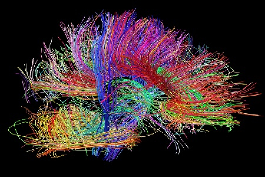
This PhD course provides a systematic and comprehensive overview of the structure and function of distinct anatomic structures (e.g. neocortex, allocortex, white matter, basal ganglia, limbic system, cerebellum, spinal cord) and how these structures connect in functional networks.
The students will be introduced to the various MRI modalities and learn how these methods provide insights into the structural and functional architecture of brain systems and how to apply their anatomical knowledge when interpreting structural and functional MR images of the human brain. The course program will also provide illustrative examples of how MRI demonstrates pathological changes in structure and function related to brain diseases, and include computer exercises on human neuroanatomy.
Linking magnetic resonance imaging (MRI) to the neuroanatomy of the human brain (“MRI neuroanatomy course”)
Attending this course, the participants will
a) acquire detailed knowledge about the main anatomic structures of the brain and their anatomical connections
b) understand the basic principles behind the major MR imaging techniques that are used to study brain structure and connectivity
c) learn how brain structure and structural connectivity can be studied with MRI techniques
d) learn how functional segregation (specialization) and functional integration interact and can be studied with MRI techniques
e) learn how MRI can be used to detect structural and functional abnormalities and what these "pathological" findings can tell us about normal brain function and structure
f) get hands-on experience on locating main anatomical structures and landmarks of the human brain through inter-active computer exercises
The course will consist of introductory lectures in the morning (four to five lectures per day), followed by supervised practical exercises based on MR imaging techniques.
PhD students (medicine, neuroscience, psychology, engineering, humanities) interested in neuroimaging. No specific qualifications required.
English
Lectures in the morning and work shops with practical exercises in small groups in the afternoon.
Ellen Garde (MR-department, Hvidovre Hospital)
Kathrine Skak Madsen (MR-department, Hvidovre Hospital)
William F.C. Baaré (MR-department, Hvidovre Hospital
Prof. Hartwig R. Siebner (MR-department, Hvidovre Hospital)
The following faculty has already confirmed their participation:
Tarek Yousry, Hartwig Siebner, Lars Hanson, Oliver Hulme, Tim Dyrby, William Baaré, Kathrine Skak Madsen, Ellen Garde, Kerstin von Plessen, Tomas Paus, Giorgio Innocenti, Katrin Amunts, Narender Ramnani.